Back Of Skull Anatomy / The Skull Anatomy And Physiology. The bone that rests on top of your spine the occipital bone is a bone that covers the back of your head; Frontal, sphenoid, ethmoid, occipital, parietal and temporal. It forms the axial skeleton together with the skull and rib cage. In the adult, the skull consists of 22 individual bones, 21 of which are immobile and united into a single unit. In the center of this region is the.
Massaging the sinus pressure points at the back of the head. Bumps on this part of the body can be hard or soft, and. The temporal bone connects to the occipital bone in the back. 1 also, some people are more prone to headaches than others. It is located next to five of the cranium bones.
Occipital Artery Anatomy Function And Significance from www.verywellhealth.com They are attached at the back of the head, and usually give rise for a headache. Massaging the sinus pressure points at the back of the head. The occipital bone is located at the back of the skull and protects the underlying cerebellum, brainstem, and occipital lobe of the cerebrum. The occipital bone is the only bone in your head that connects with your cervical spine (neck). Nerves and vessels form neurovascular bundles, which may be harmed at the sites of skull openings by pathological process or trauma. The occipital bone (/ ˌɒkˈsɪpɪtəl /) is a cranial dermal bone and the main bone of the occiput (back and lower part of the skull). Back of the head muscle structure and nerve system diagram back of the head muscle structure and nerve system diagram, vector illustration labeled medical health care scheme. The major sutures are the coronal suture, sagittal suture, lambdoid suture and squamosal sutures.
Back of the head muscle structure and nerve system diagram back of the head muscle structure and nerve system diagram, vector illustration labeled medical health care scheme.
In the center of this region is the. Overview, anterior skull base, middle skull base march 18, 2017. The occipital bone houses the back part of the brain and is one of seven bones that come together to form the skull. Axial muscles of the head, neck, and back. This portion of the skull base consists of the orbital portion of the frontal bone. Artificial human skeleton model on background, back view. The pressure points will help in dealing with the pressure points with the improvement of circulation, tension relief, and endorphins stimulation. See anatomy of the head and neck stock video clips. The skull base is the inferior portion of the neurocranium. The greater portion of the anterior floor is convex and grooved by the frontal lobe gyri. Back of the head muscle structure and nerve system diagram back of the head muscle structure and nerve system diagram, vector illustration labeled medical health care scheme. It is formed by a chain of 33 interconnected vertebrae and their intervening joints. It is also known as the calvarium.
Human anatomy for muscle, reproductive, and skeleton. The vertebral column (spine) is the bony core of the back. Bone of back of skull, find out more about bone of back of skull. This usually stems from tension in the muscles in the neck. The occipital bone overlies the occipital lobes of the cerebrum.
Anatomy The Human Skull Youtube from i.ytimg.com The vertebral column (spine) is the bony core of the back. Numerous muscles, ligaments and tendons support the spine, providing it with flexibility and a great range of motion. The skull has a single occipital condyle.7 the skull consists of five major bones: It is also known as the calvarium. Human anatomy for muscle, reproductive, and skeleton. It is trapezoidal in shape and curved on itself like a shallow dish. Artificial human skeleton model on white, back view. Back of skull anatomy :
Back of skull anatomy :
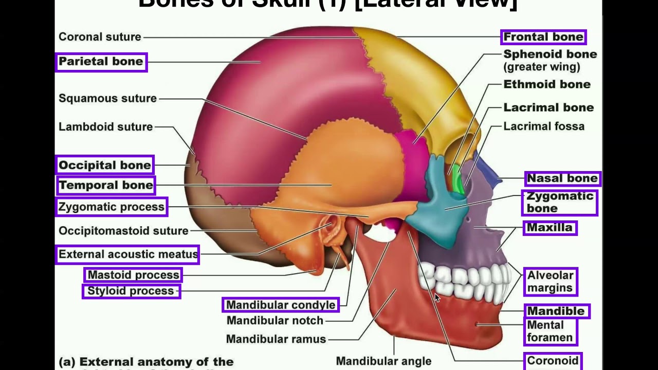
It is made up of more than 100 billion nerves that communicate in trillions of connections called synapses. The neurocranium (cranial vault) and the viscerocranium (facial skeleton). It is also known as the calvarium. Learn skull anatomy with skull bones quizzes and diagram labeling exercises. The 22nd bone is the mandible (lower jaw), which is the only moveable bone of the skull. They are attached at the back of the head, and usually give rise for a headache. The major sutures are the coronal suture, sagittal suture, lambdoid suture and squamosal sutures. The ethmoid bone forms the central part of the floor, which is the deepest area of the anterior cranial fossa. Muscle head anatomy vocal organ diagram female neck anatomy neck wireframe head neck human anatomy head artery anatomy face pharynx vector neck degree head anatomy 3d. The greater portion of the anterior floor is convex and grooved by the frontal lobe gyri. Back of the head muscle structure and nerve system diagram back of the head muscle structure and nerve system diagram, vector illustration labeled medical health care scheme. There are several important features to know about the occipital bone. See anatomy of the head and neck stock video clips.
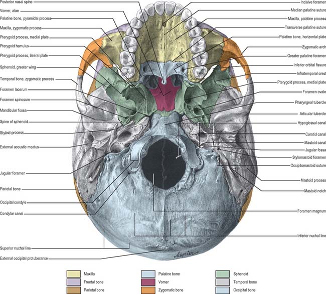
The major sutures are the coronal suture, sagittal suture, lambdoid suture and squamosal sutures. The skull base is the inferior portion of the neurocranium. The occipital bone is the only bone in your head that connects with your cervical spine (neck). A thorough description is beyond the. The head rests on the top part of the vertebral column, with the skull joining at c1.
External Skull Clinical Gate from clinicalgate.com Anterior fossa, middle fossa, and posterior fossa. There are several important features to know about the occipital bone. The eight major bones of the cranium are connected by cranial sutures, which are fibrous bands of tissue that. The pressure points will help in dealing with the pressure points with the improvement of circulation, tension relief, and endorphins stimulation. Learn about the anatomy of the skull bones and sutures as seen on ct images of the brain. See human skull anatomy stock video clips. Axial muscles of the head, neck, and back. It is also known as the calvarium.
Pain in the back of your head at the base of your skull can cause your head to hurt with dull nagging persistent pains.
It is formed by a chain of 33 interconnected vertebrae and their intervening joints. The skull base is the inferior portion of the neurocranium. In the center of this region is the. The skull is a strong, bony capsule that rests on the neck and encloses the brain. The eight major bones of the cranium are connected by cranial sutures, which are fibrous bands of tissue that. • it has the supraorbital foramen, where the supraorbital the paired parietal bones make up the top and lateral aspects of the cranium.overview, anterior skull base, middle skull base march 18, 2017. Numerous muscles, ligaments and tendons support the spine, providing it with flexibility and a great range of motion. Bumps on this part of the body can be hard or soft, and. The skull has a single occipital condyle.7 the skull consists of five major bones: From an anatomical perspective, the skull is divided into two parts: The occipital bone (/ ˌɒkˈsɪpɪtəl /) is a cranial dermal bone and the main bone of the occiput (back and lower part of the skull). The pressure points will help in dealing with the pressure points with the improvement of circulation, tension relief, and endorphins stimulation. The bone that rests on top of your spine the occipital bone is a bone that covers the back of your head;
:max_bytes(150000):strip_icc()/human-skull-with-veins-and-arteries--rear-view--1174640349-490cb7f8593945c4b1690b152e6a4074.jpg)